For the millions of people diagnosed with heart valve disease every year, there are a few options for treatment. These include open heart surgery, but some people are considered too high-risk for such a procedure. For these people, transcatheter aortic valve replacement (TAVR) has become the go-to treatment. This less-invasive procedure involves inserting a prosthetic valve to replace the damaged one, but there are still complications that can occur. If the valve doesn’t fit perfectly, it can leak. Paravalvular leak (PVL) can lead to higher mortality rates among these patients, and doctors and researchers are working to find ways to prevent this from happening.
One way to keep PVL from occurring is to make sure that the patient has a valve that fits. This can be difficult as there have been a limited number of sizes in which the valves are available, but some medical professionals have begun using 3D printing and modelling to better customize the procedure to the patient. A new study has been published that examines the effectiveness in using these technologies to treat patients.
“Right now, we rely on valve technology to assist us. But it would be nice to know what to expect, before we go in, to help us with valve selection.”
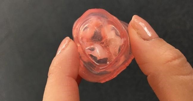
The study examined six patients who had developed PVL after TAVR for severe calcific aortic stenosis. The researchers used pre-procedure CT scans to create 3D printed models of each patient’s aortic root. The CT scans allowed them to see the location of the calcium build-up, while the 3D printed models allowed them to further evaluate the poorly fitting valves. The 3D printed models were implanted with the valves the patients had received; five patients received a 26 mm valve while one received a 23 mm valve.
The 3D printed models were then re-scanned with CT, and the images were compared with echocardiograms taken from the patients after TAVR. In every case, the fit of the 3D printed model predicted the leak seen on echo, suggesting that if the 3D modelling had been done prior to the initial procedure, the leakage could potentially have been prevented.
The study confirms that 3D modeming and 3D printing are effective ways to personalize valve size, location and placement to lower calcium build-up and prevent leaks.
Dr. Gurevich and his colleagues are working on building a library of aortic valve geometries that will be analysed by computational flow dynamics, in hopes of quantifying the amount of leak anticipated. Once enough data has been compiled, most pre-TAVR modelling can probably be done on a computer, he said, without even requiring 3D printed models. According to Dr. Gurevich, this research may even lead to the development of custom aortic valves for a perfect, patient-specific fit without risk of leakage.
Dr. Gurevich also said that it may be possible to use this type of personalized approach for other structural interventions, including left atrial appendage closure devices and mitral valve
PUT US TO THE TEST!
Let us show you want we can do!
